


Biomechanics Assessment/Gait analysis
A biomechanical assessment involves a detailed examination of the lower limbs, focusing on structure, alignment, strengths, and weaknesses. This assessment looks at the pelvis, legs, knees, and feet as a whole, as these areas are closely connected and pain in one region may stem from a weakness or structural issue elsewhere.
The foot is a complex structure made up of 28 bones, 214 ligaments, and 38 muscles, all working together to support body weight during everyday movement.
A biomechanical assessment is particularly valuable if you are experiencing foot or lower-limb pain without a clear cause identified through standard interventions. It provides a starting point for understanding the source of your symptoms, determining appropriate treatment, and identifying whether further investigations are required..
What happens during a biomechanical assessment?
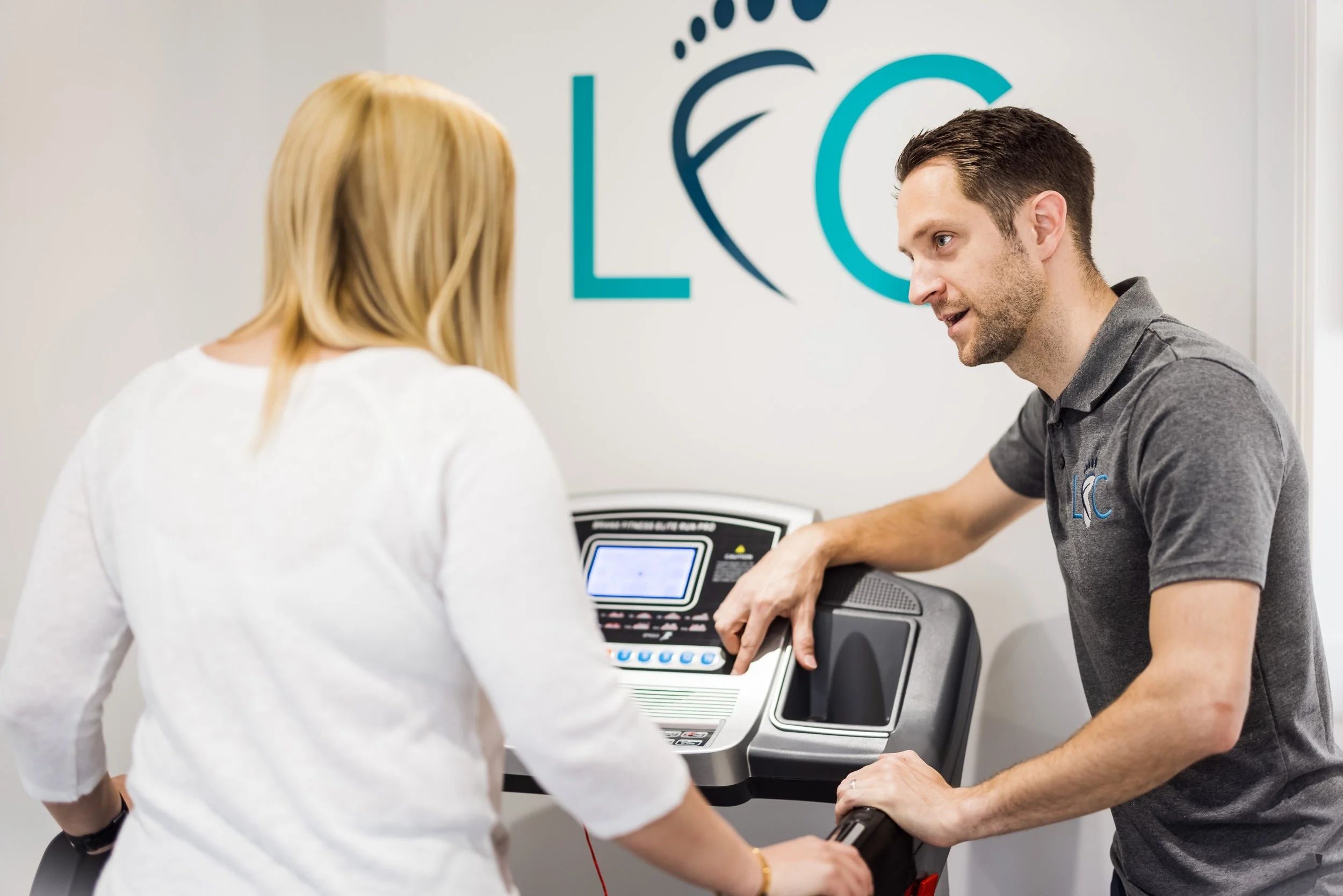
During the assessment, we will examine your gait, walking pattern, and overall movement. Depending on when and how your symptoms occur, this may include using a treadmill or pressure plate. These types of assessments are carried out for all individuals, from everyday walkers experiencing pain through to top class athletes to help guide an appropriate intervention.
A range of treatment options may be recommended following a biomechanical assessment, depending on your results. For individuals with good structural foot mechanics, the podiatrist may simply advise on suitable footwear to help reduce the risk of future problems. Small changes—such as choosing trainers designed for your gait or using simple insoles—can be highly effective.
If your podiatrist identifies that your foot mechanics are contributing to your pain or injury, prescription insoles or custom-made orthotics may be recommended, along with exercises to improve muscle strength and flexibility.
Referral to other healthcare professionals, such as your GP, physiotherapist, or rheumatologist, may also be necessary. In some cases, further imaging—such as X-rays, MRIs, or CT scans—may be advised.


-
An ‘orthosis’ is a medical term which describes a device that supports, realigns or assists in the function of the musculo-skeletal system. In podiatry we use foot orthoses, or commonly known as orthotics. Therefore “foot orthoses” are designed to support, align or improve the function of the feet and lower limbs during gait.
Functional orthotics apply forces to the feet enabling the podiatrist to alter certain movements or off-load stress. Their prescription is based on biomechanical and anatomical assessments but may also involve a degree of lateral thinking by the podiatrist if you have complex problems.
Non-functional orthotics may be designed to improve skin and tissue viability or off-load painful pressure areas.
-
Painful pressure areas can be identfied from the wear and wear on your feet. If we require more detail we use a medicapteur pressure plate system. The impressions are scanned into a computer system, which highlights areas of the foot experiencing excessive or insufficient pressure. This information helps guide treatment, whether that involves joint mobilisation, taping, or prescribing custom-made or off-the-shelf orthotics tailored to the specific needs of each foot.
-
Due to advances in technology, we’re now able to offer the best 3D scanned orthotics with our scanning equipment in clinic
-
A biomechanics appointment can help with a range of foot conditions including:
Achilles tendinopathy
Ankle impingement
Big toe joint pain/bunion pain
Bursitis/capsulitis
Plantar Fasciitis/heel pain
metatarsalgia/forefoot pain
Flat feet
Pes planus
Arthritis
Ankle sprains
Mortons Neuroma/nerve pain
Tibialis posterior and anterior pain
-
Book an appointment or call 01202 721417 to speak to a knowledgeable health care professional.